Bones In Leg Diagram - Calf Anatomy - The tibia, commonly known as the 'shin bone', is the largest and most medial of the two.. Inflammation of navicular bone and/or bursa. Inside of arm muscle and bone 12 photos of the inside of arm muscle and bone , bone The human leg consists of 8 bones, 4 per leg. The lower leg is comprised of two bones the tibia and the smaller fibula. Related posts of bones leg diagram picture bones and wrist and hand palmar view.
Related posts of bones leg diagram picture bones and wrist and hand palmar view. The ilium is the bone at the top of the waist, while the pubis bones are found just below the ilium. Quadriceps and hamstring muscles are the major ones which are associated with the knee joint. Inflammation of navicular bone and/or bursa. The human leg consists of 8 bones, 4 per leg.
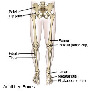
Knee Anatomy from embed.widencdn.net Numbered one through five the bone that sits behind the big toe is no. This diagram depicts diagram leg bones anatomy.human anatomy diagrams show internal organs, cells, systems, conditions, symptoms and sickness information and/or tips for healthy living. The second metatarsal bone is the longest. The human leg consists of 8 bones, 4 per leg. The ischium is located just behind the pubis bone. Most of the leg skeleton has bony prominences and margins that can be palpated and some serve as anatomical landmarks that define the extent of the leg. The femur, or thighbone, is the longest and largest bone in the human body. At the distal end of the femur, two rounded condyles meet the tibia and fibula bones of the lower leg to form the knee joint.
The ligament joining the two bones of the lower leg (tibia and fibula), called the syndesmotic ligament, is injured.
Click now to learn more about the bones, muscles, and soft tissues tibia: Its decrease finish helps create the knee joint. Blood vessels and nerves enter the bone through the nutrient foramen. This long bone connects with the knee at one end and the ankle at the other. They are numbered from one to five, starting from the medial (inner) side of the foot. Four quadriceps muscles are present in front of the knee which help in straightening the leg from the knee. Bone diagram forehead (frontal bone) nose bones (nasals) cheek bone (zygoma) upper jaw (maxilla) lower jaw (mandible) breast bone (sternum) upper arm bone (humerus) lower arm bone (ulna) thigh bone (femur) collar bone (clavicle) toe bones (phalanges) ankle bones (tarsals) kneecap (patella) shin bone Cancellous bone produces red blood cells, platelets, and white blood cells. This area is commonly referred to as the calf. The second largest bone in body is the tibia, also called the shinbone. The foot bones shown in this diagram are the talus, navicular, cuneiform, cuboid, metatarsals and. Inside of arm muscle and bone 12 photos of the inside of arm muscle and bone , bone The major bones of the leg are the femur (thigh bone), tibia (shin bone), and adjacent fibula, and these are all long bones.
These bones are firmly attached with the help of muscles and tendons which also save the bones from injury. The medial, larger bone of the lower leg. Four quadriceps muscles are present in front of the knee which help in straightening the leg from the knee. The proximal portion of the tibia is tibial plateau which acts as a cusp for the knee, the distal portion tapers into the medial malleoli and the concave surface which articulates with the talus at the ankle joint. A high ankle sprain causes pain and swelling similar to a.
The Bones Of The Leg Stock Image F001 7220 Science Photo Library from media.sciencephoto.com The five metatarsals are the long bones that link the tarsal bones to the toes, seen in yellow in the diagram below. Bones and wrist and hand palmar view 12 photos of the bones and wrist and hand palmar view bones of the wrist and hand palmar view, bone, bones of the wrist and hand palmar view. Hip and leg bone diagram : The ischium is located just behind the pubis bone. Master leg and knee anatomy using our topic page. Distal end of right humerus. Leg bone diagram / bones of the human leg 17. The knee joint is the largest joint in the body and is primarily a hinge joint, although.
Master leg and knee anatomy using our topic page.
The second metatarsal bone is the longest. Related posts of bones leg diagram picture. Four quadriceps muscles are present in front of the knee which help in straightening the leg from the knee. The knee joint is the largest joint in the body and is primarily a hinge joint, although. This long bone connects with the knee at one end and the ankle at the other. Schema de legs bones diagram diagram showing bones inside human leg ready to jump stock file skeleton of a cat diagram ver 2 svg disposition of rotator cuff muscles diagram. Next to the tibia is the fibula, the thinner,. The foot bones shown in this diagram are the talus, navicular, cuneiform, cuboid, metatarsals and. Performance horses tend to suffer from this degenerative disease. The pubis curves downward and forwards from the ileum. The smaller lateral bone of the lower leg. The femur, or thighbone, is the longest and largest bone within the human physique. The femur, or thighbone, is the longest and largest bone in the human body.
Cancellous bone produces red blood cells, platelets, and white blood cells. The femur, or thighbone, is the longest and largest bone within the human physique. The ilium is the bone at the top of the waist, while the pubis bones are found just below the ilium. The lower leg extends from the knee to the ankle. They are numbered from one to five, starting from the medial (inner) side of the foot.
Leg Fracture What You Need To Know from www.drugs.com Most of the leg skeleton has bony prominences and margins that can be palpated and some serve as anatomical landmarks that define the extent of the leg. Related posts of bones leg diagram picture. Bones of right thigh and leg. The ischium is located just behind the pubis bone. The femur, or thighbone, is the longest and largest bone within the human physique. Inside of arm muscle and bone 12 photos of the inside of arm muscle and bone , bone Blood vessels and nerves enter the bone through the nutrient foramen. You can palpate its anterior border when you run your finger down the anterior aspect of your leg.
Master leg and knee anatomy using our topic page.
The knee is a strong but flexible hinge joint that uses muscles and ligaments to withstand the torques and strains of powerful leg movements. He leg's main function in the human is for locomotion and support of the rest of the body. Click now to learn more about the bones, muscles, and soft tissues tibia: This diagram depicts diagram leg bones anatomy.human anatomy diagrams show internal organs, cells, systems, conditions, symptoms and sickness information and/or tips for healthy living. At the distal end of the femur, two rounded condyles meet the tibia and fibula bones of the lower leg to form the knee joint. The bones of the leg are the femur, tibia, fibula and patella.the foot bones shown in this diagram are the talus, navicular, cuneiform, cuboid, metatarsals and calcaneus. The thigh bone, or femur, is the large upper leg bone that connects the lower leg bones (knee joint) to the pelvic bone (hip joint). The largest and most medial leg bone, forming both the knee and ankle joints. Pin on medical websites we like. The lower leg is comprised of two bones the tibia and the smaller fibula. The foot bones shown in this diagram are the talus, navicular, cuneiform, cuboid, metatarsals and. Distal end of right humerus. The lower leg is comprised of two bones, the tibia and the smaller fibula.


0 Komentar